Toggle light / dark theme

Results of a study show convergent genetic adaptation under hypoxia (lack of oxygen) between populations living at high-altitude in the Himalayan region such as Tibetans and Sherpas, and the development of oxygen-starved cancer cells. The study was directed by Rodrigo Toledo, Head of the Vall d’Hebron Institute of Oncology’s (VHIO) Biomarkers and Clonal Dynamics Group and published in the journal Cancer Discovery.

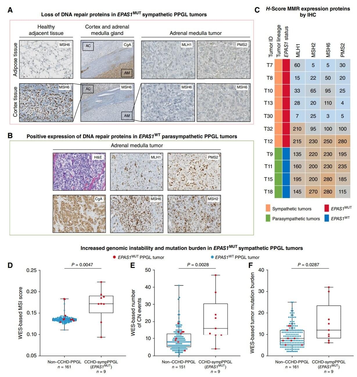
Patients with cyanotic congenital heart disease (CCHD) are chronically hypoxic and have an estimated six-fold higher risk of developing pheochromocytoma and paraganglioma (PPGL), which are associated with (NETs) of the adrenal glands and/or paraganglia, respectively. These cancers can continue to grow and proliferate under chronic hypoxia.

“With this study, we aimed to achieve deeper insights into how tumors can survive, grow, and even metastasize under low oxygen conditions, known as hypoxia. Our findings reveal a broad convergence in in tumors that continue to develop and grow under hypoxia, and in high-altitude populations who thrive in such a challenging environment,” said Toledo, corresponding author of this present article.

A research team from Helmholtz Munich and the Technical University of Munich has developed an advanced delivery system that transports gene-editing tools based on the CRISPR/Cas9 gene-editing system into living cells with significantly greater efficiency than before. Their technology, ENVLPE, uses engineered non-infectious virus-like particles to precisely correct defective genes—demonstrated successfully in living mouse models that are blind due to a mutation.

This system also holds promise for advancing by enabling precise genetic manipulation of engineered , making them more universally compatible and thus more accessible for a larger group of cancer patients.

The work is published in the journal Cell.

While it may be an unfamiliar sensation to humans, electroreception is relatively commonplace in the animal kingdom. Sharks, bees and even the platypus all share this ability to detect electric fields in their environment.

Scientists at UC Santa Barbara have just added to that list. A team of researchers led by Matthieu Louis found that fruit fly larvae can sense electric fields and navigate toward the negative electric potential using a small set of sensory neurons in their head.

The findings, published in Current Biology, present an immense opportunity. Fruit flies are arguably the most commonly used experimental animals, the basis for studies in fields as disparate as genetics, neurobiology and aging. Uncovering electroreception in fruit flies opens new avenues of research into the basis of this sense and could even lead to new techniques in bioengineering.

Microbial life has dominated Earth’s history but left a sparse fossil record, greatly hindering our understanding of evolution in deep time. However, bacterial metabolism has left signatures in the geochemical record, most conspicuously the Great Oxidation Event (GOE). We combine machine learning and phylogenetic reconciliation to infer ancestral bacterial transitions to aerobic lifestyles, linking them to the GOE to calibrate the bacterial time tree. Extant bacterial phyla trace their diversity to the Archaean and Proterozoic, and bacterial families prior to the Phanerozoic. We infer that most bacterial phyla were ancestrally anaerobic and adopted aerobic lifestyles after the GOE. However, in the cyanobacterial ancestor, aerobic metabolism likely predated the GOE, which may have facilitated the evolution of oxygenic photosynthesis.

First, the team discovered that heparan sulfate (HSPG), a sulfated glycoprotein on the cell surface, plays a crucial role in attracting LNPs and facilitating mRNA entry into the cell.

- Second, they identified V-ATPase, a proton pump at the endosome, which acidifies the vesicle and causes LNPs to become positively charged, enabling them to temporarily disrupt the endosomal membrane and release the mRNA into the cytoplasm to be expressed.

- Lastly, the study uncovered the role of TRIM25, a protein involved in the cellular defense mechanism. TRIM25 binds to and induces the rapid degradation of exogenous mRNAs, preventing their function.

So how do the mRNA vaccines evade this cellular defense? A key finding of the study was that mRNA molecules containing a special modification called N1-methylpseudouridine (m1Ψ)—which was awarded the 2023 Nobel Prize in Physiology or Medicine—can evade TRIM25 detection. This modification prevents TRIM25 from binding to mRNA, enhancing the stability and effectiveness of mRNA vaccines. This discovery not only explains how mRNA vaccines evade cellular surveillance mechanisms but also emphasizes the importance of this modification in enhancing the therapeutic potential of mRNA-based treatments.

Additionally, the research highlighted the critical role of proton ions in this process. When the LNPs rupture the endosomal membrane, proton ions are released into the cytoplasm, which activates TRIM25. These proton ions act as a signal that alerts the cell to the invading foreign RNA, which in turn triggers a defense response. This is the first study to demonstrate that proton ions serve as immune signaling molecules, providing new insights into how cells protect themselves from foreign RNA.


A team of researchers has uncovered a key cellular mechanism that affects the function of mRNA vaccines and therapeutics. Their study, recently published in Science, provides the first comprehensive understanding of how mRNA vaccines are delivered, processed, and degraded within cells—a breakthrough that could pave the way for more effective vaccines and RNA-based treatments.

Liver regeneration after hepatectomy follows accurate coordination with the body’s specific requirements1–3. However, the molecular mechanisms, factors and particular hepatocyte population influencing its efficiency remain unclear. Here we report on a unique regeneration mechanism involving unconventional RPB5 prefoldin interactor 1 (URI1), which exclusively colocalizes with, binds to and activates glutamine synthase (GS) in pericentral hepatocytes. Genetic GS or URI1 depletion in mouse pericentral hepatocytes increases circulating glutamate levels, accelerating liver regeneration after two-third hepatectomy. Conversely, mouse hepatocytic URI1 overexpression hinders liver restoration, which can be reversed by elevating glutamate through supplementation or genetic GS depletion. Glutamate metabolically reprograms bone-marrow-derived macrophages, stabilizing HIF1α, which transcriptionally activates WNT3 to promote YAP1-dependent hepatocyte proliferation, boosting liver regeneration. GS regulation by URI1 is a mechanism that maintains optimal glutamate levels, probably to spatiotemporally fine-tune liver growth in accordance with the body’s homeostasis and nutrient supply. Accordingly, in acute and chronic injury models, including in cirrhotic mice with low glutamate levels and in early mortality after liver resection, as well as in mice undergoing 90% hepatectomy, glutamate addition enhances hepatocyte proliferation and survival. Furthermore, URI1 and GS expression co-localize in human hepatocytes and correlate with WNT3 in immune cells across liver disease stages. Glutamate supplementation may therefore support liver regeneration, benefiting patients awaiting transplants or recovering from hepatectomy.

© 2025. The Author(s), under exclusive licence to Springer Nature Limited.


Damage to the mitochondria, the “power plants” of the cells, contributes to many diseases. Researchers from Heinrich Heine University Düsseldorf (HHU) and the University of Cologne led by HHU professor of medicine Dr David Pla-Martín, now describe in the scientific journal Science Advances how cells with defective mitochondria activate a special recycling system to eliminate damaged genetic material.

Damage to the genetic material of mitochondria – the mitochondrial DNA or mtDNA for short – can lead to diseases such as Parkinson’s, Alzheimer’s, amyotrophic lateral sclerosis (ALS), cardiovascular diseases and type 2 diabetes. Such damage also speeds up the ageing process. However, the cells are normally capable of identifying such damage and reacting.

Damage to the genetic material of mitochondria—the mitochondrial DNA or mtDNA for short—can lead to diseases such as Parkinson’s, Alzheimer’s, amyotrophic lateral sclerosis (ALS), cardiovascular diseases and type 2 diabetes. Such damage also speeds up the aging process. However, the cells are normally capable of identifying such damage and reacting.

Scientists from University Hospital Düsseldorf and HHU have—in collaboration with the University of Cologne and the Center for Molecular Medicine Cologne (CMMC)—discovered a mechanism which protects and repairs the mitochondria. The research team, headed by Professor Pla-Martín from the Institute of Biochemistry and Molecular Biology I at HHU, has identified a specialized recycling system, which cells activate when they identify damage to the mtDNA.

According to the authors in Science Advances, this mechanism relies on a known as retromer and the lysosomes—cell organelles containing digestive enzymes. These special cellular compartments act like recycling centers, eliminating the damaged genetic material.

Constructed strain achieves record-high yield from methanol, advancing ecofriendly biomanufacturing. Researchers from Osaka Metropolitan University have discovered the ideal genetic “recipe” to turn yeast into a tiny yet powerful eco-friendly factory that converts methanol into D-lactic acid, a key compound used in biodegradable plastics and pharmaceuticals.

This approach could help reduce reliance on petroleum-based processes and contribute to more sustainable chemical production.

Lactic acid is widely used in food, cosmetics, pharmaceuticals and bioplastics.