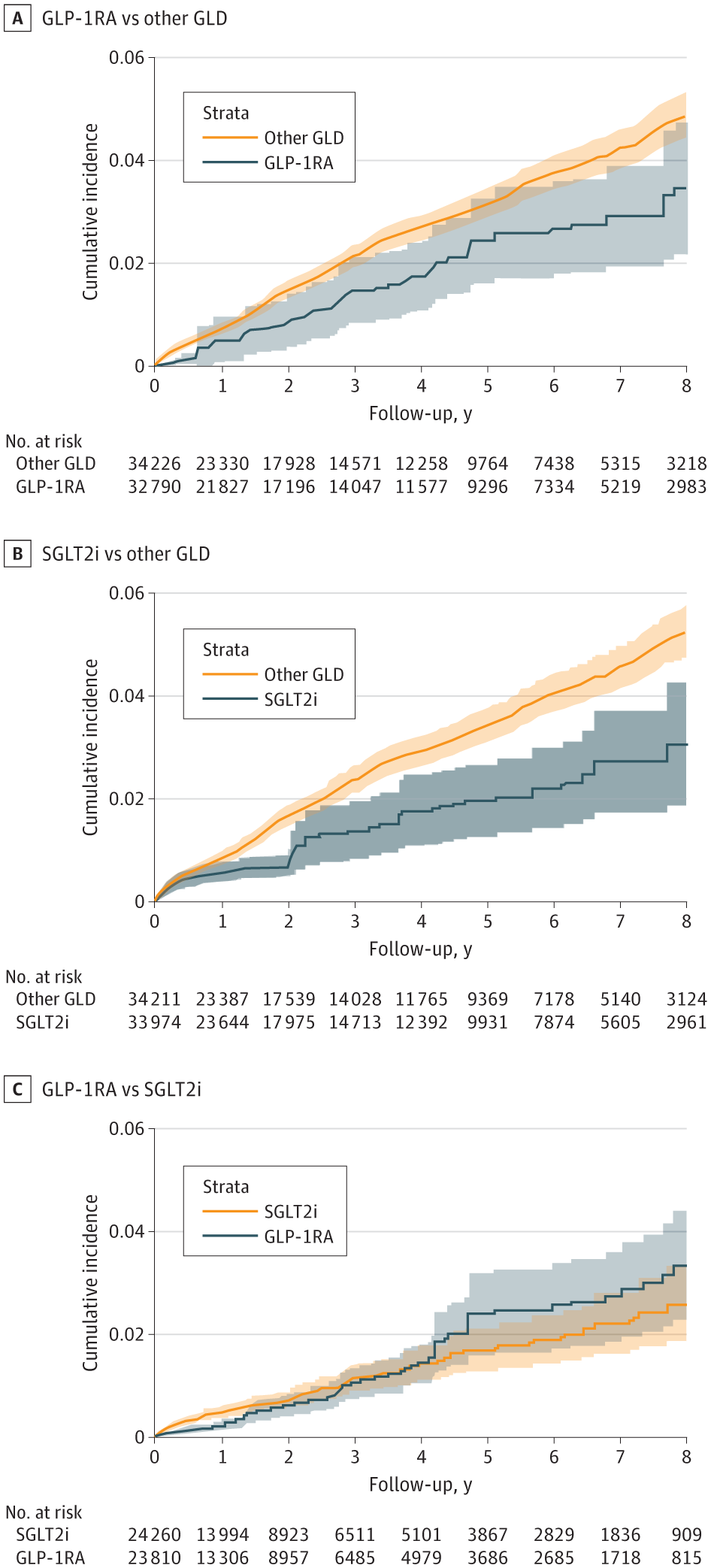
This population-based cohort study examines the association of glucagon-like peptide-1 receptor agonists and sodium-glucose cotransporter-2 inhibitors with the risk of Alzheimer disease and related dementias in patients with type 2 diabetes.
This population-based cohort study examines the association of glucagon-like peptide-1 receptor agonists and sodium-glucose cotransporter-2 inhibitors with the risk of Alzheimer disease and related dementias in patients with type 2 diabetes.
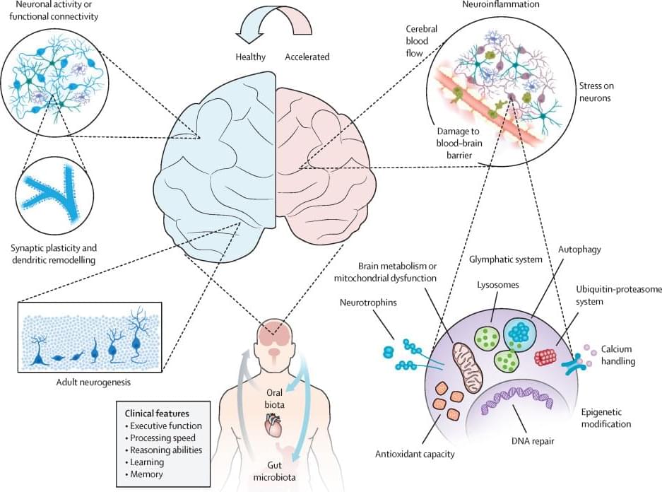
Ageing is a scientifically fascinating and complex biological occurrence characterised by morphological and functional changes due to accumulated molecular and cellular damage impairing tissue and organ function. Ageing is often accompanied by cognitive decline but is also the biggest known risk factor for Alzheimer’s disease, the most common form of dementia. Emerging evidence suggests that sedentary and unhealthy lifestyles accelerate brain ageing, while regular physical activity, high cardiorespiratory fitness (CRF), or a combination of both, can mitigate cognitive impairment and reduce dementia risk.
17K likes, — vaibhavsisinty on March 27, 2025: “Your Future Kids Might Be Genetically Engineered🤯… [genetic engineering, CRISPR, designer babies, IVF, in vitro gametogenesis, gene editing, human evolution, bioethics, futuristic science, AI in healthcare, medical advancements, artificial reproduction, skin cell gametes, future tech, DNA modification, biotechnology]”
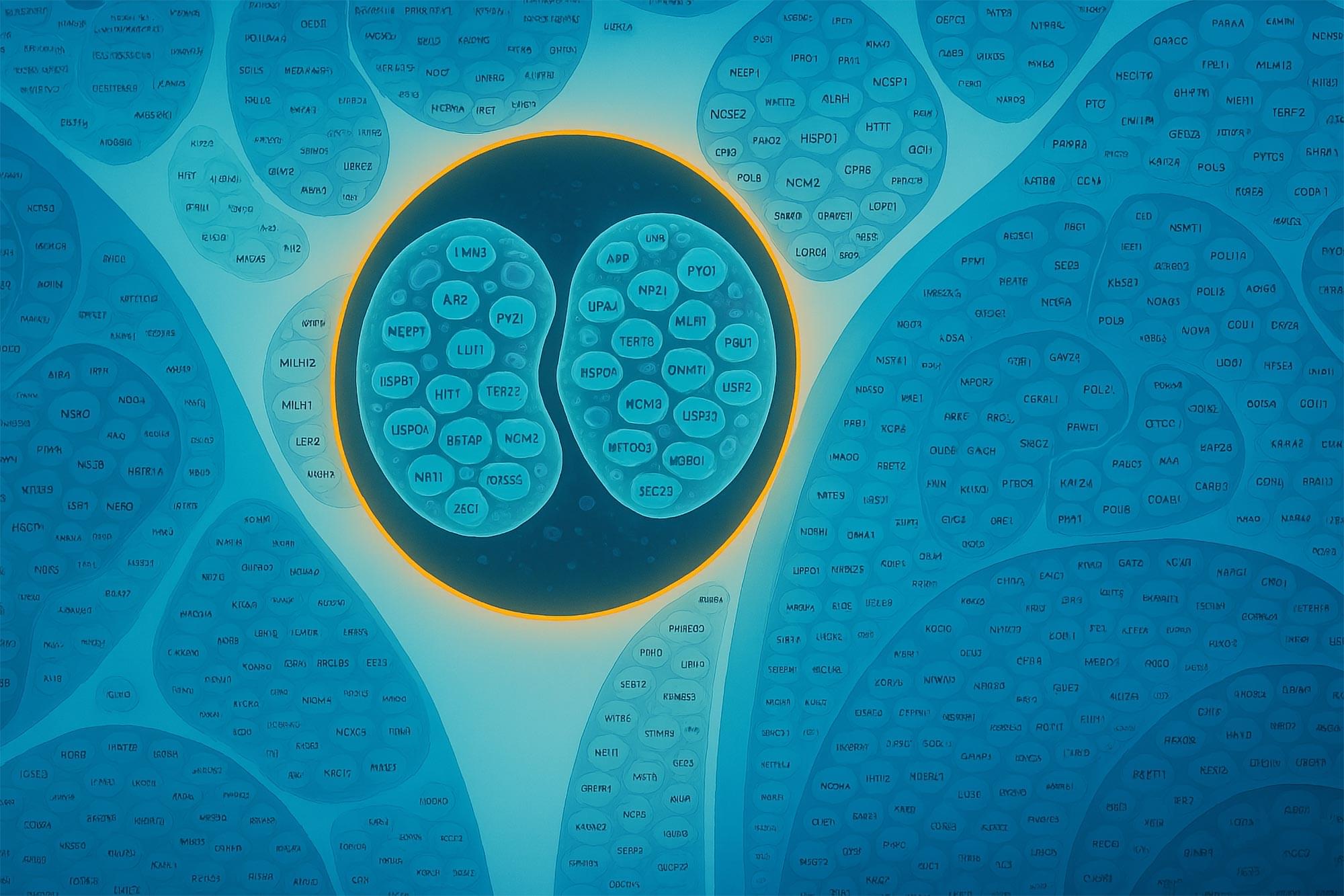
For the first time, scientists have built a detailed, interactive map of a human cell, revealing how thousands of proteins organize and work together.
Using advanced imaging and AI tools like GPT-4, they uncovered hundreds of previously unknown protein functions and identified key cellular assemblies tied to childhood cancers. This map not only changes how we study cell biology but could also transform our understanding of disease at the molecular level.
Mapping the Human Cell: A 400-Year Quest.
Posted in biotech/medical, health | Leave a Comment on Moving forward from Cockcroft-Gault creatinine clearance to race-free estimated glomerular filtration rate to improve medication-related decision-making in adults across healthcare settings: A consensus of the National Kidney Foundation Workgroup for Implementation of Race-Free eGFR-Based Medication-Related Decisions
![]()
The NKF Workgroup for Implementation of Race-Free eGFR-Based Medication-Related Decisions suggests that health systems, health settings, clinical laboratories, electronic health record systems, compendia and data vendors, and healthcare practitioners involved with medication-related decision-making …
Lead author Joseph Silk, a professor in the Department of Physics and Astronomy at Johns Hopkins University, explained that this discovery could change our understanding of how galaxies formed. We know these monster black holes exist at the center of galaxies near our Milky Way, but the big surprise now is that they were present at the beginning of the universe as well and were almost like building blocks or seeds for early galaxies.
The study, published in the Astrophysical Journal Letters, analyzed distant galaxies from the early universe observed through the Webb telescope. These galaxies appeared much brighter than expected and contained unusually high numbers of young stars and supermassive black holes.
The findings challenge the conventional idea that black holes formed after the collapse of supermassive stars and that galaxies formed after the first stars appeared. Instead, the analysis suggests that black holes and galaxies coexisted and influenced each other’s development during the first 100 million years of the universe.
Researchers from the University of Colorado Cancer Center have solved a cellular mystery that may lead to better therapies for colorectal and other types of cancer.
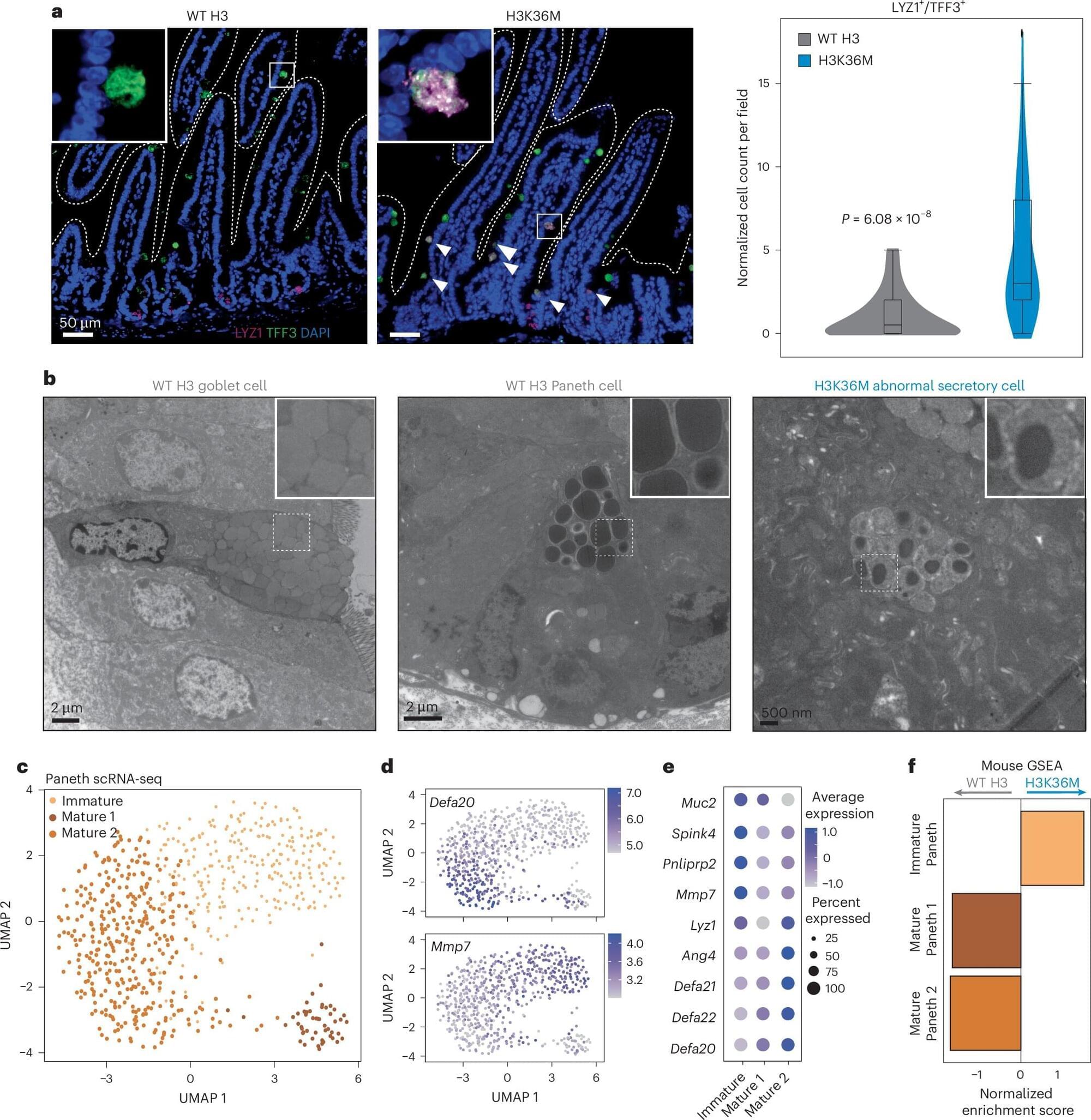
Peter Dempsey, Ph.D., professor of pediatrics– developmental biology in the CU School of Medicine, and Justin Brumbaugh, Ph.D., assistant professor of molecular, cellular, and developmental biology at CU Boulder, recently published a paper in the journal Nature Cell Biology showing the importance of the H3K36 methylation process in regulating plasticity and regeneration in intestinal cells.
“The intestine has an enormous ability to regenerate itself after injury, and it does this through a model of dedifferentiation,” Dempsey explains. “The cells dedifferentiate back into a type of regenerative stem cell after injury, and those stem cells eventually recover the intestine and turn back to normal cells.”
Join us on Patreon! https://www.patreon.com/MichaelLustgartenPhD
Discount Links/Affiliates:
Blood testing (where I get the majority of my labs): https://www.ultalabtests.com/partners/michaellustgarten.
At-Home Metabolomics: https://www.iollo.com?ref=michael-lustgarten.
Use Code: CONQUERAGING At Checkout.
Clearly Filtered Water Filter: https://get.aspr.app/SHoPY
Epigenetic, Telomere Testing: https://trudiagnostic.com/?irclickid=U-s3Ii2r7xyIU-LSYLyQdQ6…M0&irgwc=1
Use Code: CONQUERAGING
NAD+ Quantification: https://www.jinfiniti.com/intracellular-nad-test/

Just as pilots use flight simulators to safely practice complex maneuvers, scientists may soon conduct experiments on a highly realistic simulation of the mouse brain. In a new study, researchers at Stanford Medicine and their collaborators developed an artificial intelligence.
Artificial Intelligence (AI) is a branch of computer science focused on creating systems that can perform tasks typically requiring human intelligence. These tasks include understanding natural language, recognizing patterns, solving problems, and learning from experience. AI technologies use algorithms and massive amounts of data to train models that can make decisions, automate processes, and improve over time through machine learning. The applications of AI are diverse, impacting fields such as healthcare, finance, automotive, and entertainment, fundamentally changing the way we interact with technology.
Posted in biotech/medical, evolution | Leave a Comment on Viral oncogenes, viruses, and cancer: a third-generation sequencing perspective on viral integration into the human genome

The link between viruses and cancer has intrigued scientists for decades. Certain viruses have been shown to be vital in the development of various cancers by integrating viral DNA into the host genome and activating viral oncogenes. These viruses include the Human Papillomavirus (HPV), Hepatitis B and C Viruses (HBV and HCV), Epstein-Barr Virus (EBV), and Human T-Cell Leukemia Virus (HTLV-1), which are all linked to the development of a myriad of human cancers. Third-generation sequencing technologies have revolutionized our ability to study viral integration events at unprecedented resolution in recent years. They offer long sequencing capabilities along with the ability to map viral integration sites, assess host gene expression, and track clonal evolution in cancer cells.